CUBA | Artificial intelligence and radiology in the COVID-19 battle

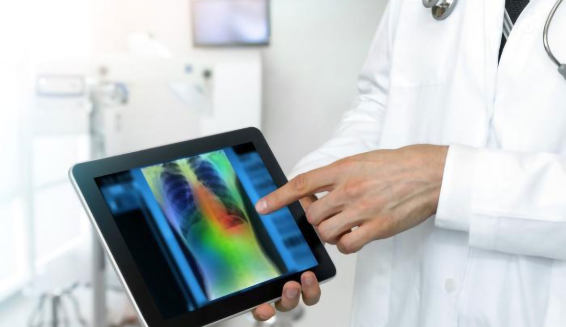
HAVANA, Cuba, January 2, 2021 - Granma - Specialists from Cuba’s Neurosciences Center (Cneuro) and the Cuban Society of Medical Imaging, have undertaken a scientific project directed toward perfecting the use of chest x-rays and Computerized Axial Tomography (CAT) in diagnosis, prognosis and follow-up of COVID-19 patients, with the use of artificial intelligence tools.
Doctor of Science Evelio Gonzalez Dalmau, head of the project and of Cneuro’s department of Magnetic Resonance Imaging and Optogenetics, told Granma that the goal is to expand the medical contributions of radiological studies through the use of artificial intelligence techniques, to improve the quality of images, provide more information and contribute to quantifying and categorizing the lesions detected with greater accuracy.
He emphasized the advantages of increasing the effectiveness of chest X-rays in the fight against the pandemic, since it is an inexpensive, rapid and widely available procedure that can contribute greatly to the early diagnosis of the disease and follow-up of hospitalized and convalescent patients.
With the project we seek to develop an automated, standardized radiological report, which will allow for the creation of a database on which the clinical histories of these patients are registered, he explained.
Dr. Gonzalez pointed out that even as the number of cases is declining significantly and the pandemic is brought under control, the project will remain valid, as it will be applicable to other pulmonary studies related, for example, to cancer, statistical follow-up of the effect of drugs and post-COVID research.
To discuss the subject, the Hermanos Ameijeiras Hospital hosted the I Virtual Conference on Imaging and COVID-19, organized by the Cuban Society of Imaging and Cneuro, with the participation of renowned professionals from different institutions across the country, as well as Spain and Argentina.
